Figure 6
Scientific Analysis of Eucharistic Miracles: Importance of a Standardization in Evaluation
Kelly Kearse* and Frank Ligaj
Published: 13 November, 2024 | Volume 8 - Issue 1 | Pages: 078-088
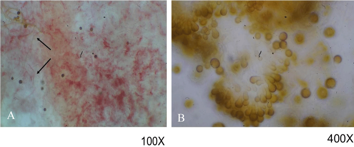
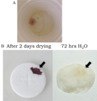
Figure 6:
Microscopic examination of reddish growths on non-consecrated wafers. Samples were examined under 100x (A) and 400x (B). Hyphae are indicated in (A) with arrows. Structures in (B) were identified as conidia and chlamydospores, similar to those seen with Epicoccum nigrum.
Read Full Article HTML DOI: 10.29328/journal.jfsr.1001068 Cite this Article Read Full Article PDF
More Images
Similar Articles
-
Awareness level on the role of forensic DNA database in criminal investigation in Nigeria: A case study of Benin cityNwawuba Stanley Udogadi*,Akpata Chinyere Blessing Nkiruka . Awareness level on the role of forensic DNA database in criminal investigation in Nigeria: A case study of Benin city. . 2020 doi: 10.29328/journal.jfsr.1001019; 4: 007-014
-
Awareness level on the relevance of forensics in criminal investigation in NigeriaOmorogbe Owen Stephen,Orhue Osazee Kelvin,Ehikhamenor Edeaghe,Nwawuba Stanley Udogadi*. Awareness level on the relevance of forensics in criminal investigation in Nigeria. . 2021 doi: 10.29328/journal.jfsr.1001028; 5: 053-057
-
Comparative analysis of mobile forensic proprietary tools: an application in forensic investigationParth Chauhan*,Tamanna Jaitly,Animesh Kumar Agrawal. Comparative analysis of mobile forensic proprietary tools: an application in forensic investigation. . 2022 doi: 10.29328/journal.jfsr.1001039; 6: 077-082
-
Forensic Analysis of WhatsApp: A Review of Techniques, Challenges, and Future DirectionsNishchal Soni*. Forensic Analysis of WhatsApp: A Review of Techniques, Challenges, and Future Directions. . 2024 doi: 10.29328/journal.jfsr.1001059; 8: 019-024
-
Analysis and Comparison of Social Media Applications Using Forensic Software on Mobile DevicesHüseyin Çakır*, Merve Hatice Karataş . Analysis and Comparison of Social Media Applications Using Forensic Software on Mobile Devices. . 2024 doi: 10.29328/journal.jfsr.1001065; 8: 058-063
-
Scientific Analysis of Eucharistic Miracles: Importance of a Standardization in EvaluationKelly Kearse*,Frank Ligaj. Scientific Analysis of Eucharistic Miracles: Importance of a Standardization in Evaluation. . 2024 doi: 10.29328/journal.jfsr.1001068; 8: 078-088
-
Biotechnology in Forensic Science: Advancements and ApplicationsSunny Antil,Vandana Joon*. Biotechnology in Forensic Science: Advancements and Applications. . 2025 doi: 10.29328/journal.jfsr.1001073; 9: 007-014
-
Digital Forensics and Media Offences – Investigate Synergy in the Cyber AgeGauri Goyal*. Digital Forensics and Media Offences – Investigate Synergy in the Cyber Age. . 2025 doi: 10.29328/journal.jfsr.1001074; 9: 015-020
Recently Viewed
-
Personal and academic factors of stress in nursing students during clinical practices in the context of COVID-19Lester Fidel García Guzmán*,Dulce Maria Oviedo Martinez,Alexis Silva López,Makorre Wilson Mohochi. Personal and academic factors of stress in nursing students during clinical practices in the context of COVID-19. Clin J Nurs Care Pract. 2022: doi: 10.29328/journal.cjncp.1001041; 6: 014-019
-
Determinants of health seeking behaviour of women with obstetric fistula in south- south and south east, Nigeria: A review of the impact of availability and quality of health care services through a cross-sectional studyPeters Grace E*,Ononokpono Dorathy N,Willson Nsikanabasi U,Oko Nnabuike I,Peters EJ. Determinants of health seeking behaviour of women with obstetric fistula in south- south and south east, Nigeria: A review of the impact of availability and quality of health care services through a cross-sectional study. Clin J Obstet Gynecol. 2021: doi: 10.29328/journal.cjog.1001088; 4: 060-065
-
Anterior Laparoscopic Approach Combined with Posterior Approach for Lumbosacral Neurolysis: A Case ReportSheng Wang#,Nan Lu,Yongchuan Li,Xiaohuang Tu*,Aimin Chen*. Anterior Laparoscopic Approach Combined with Posterior Approach for Lumbosacral Neurolysis: A Case Report. Arch Clin Exp Orthop. 2024: doi: 10.29328/journal.aceo.1001020; 8: 010-013
-
Persistent Lumbar Pain and Fever: Osteomyelitis as Diagnosis ChallengeAlicia Cárdenas García*, Sara García Mateo, María Rodríguez Pérez, José Carlos Sureda Gil, María Teresa Gómez Álvarez, Francisco de Borja Hernández Moreno, Anna de Paola Prato. Persistent Lumbar Pain and Fever: Osteomyelitis as Diagnosis Challenge. Arch Clin Exp Orthop. 2024: doi: 10.29328/journal.aceo.1001019; 8: 005-009
-
Superior Gluteal Artery Pseudoaneurysm following a Periacetabular OsteotomyTess Szekelyi, Xavier Lannes, Mouas Jammal, Salah Dine Qanadli, Michael Wettstein*. Superior Gluteal Artery Pseudoaneurysm following a Periacetabular Osteotomy. Arch Clin Exp Orthop. 2024: doi: 10.29328/journal.aceo.1001018; 8: 001-004
Most Viewed
-
Evaluation of Biostimulants Based on Recovered Protein Hydrolysates from Animal By-products as Plant Growth EnhancersH Pérez-Aguilar*, M Lacruz-Asaro, F Arán-Ais. Evaluation of Biostimulants Based on Recovered Protein Hydrolysates from Animal By-products as Plant Growth Enhancers. J Plant Sci Phytopathol. 2023 doi: 10.29328/journal.jpsp.1001104; 7: 042-047
-
Sinonasal Myxoma Extending into the Orbit in a 4-Year Old: A Case PresentationJulian A Purrinos*, Ramzi Younis. Sinonasal Myxoma Extending into the Orbit in a 4-Year Old: A Case Presentation. Arch Case Rep. 2024 doi: 10.29328/journal.acr.1001099; 8: 075-077
-
Feasibility study of magnetic sensing for detecting single-neuron action potentialsDenis Tonini,Kai Wu,Renata Saha,Jian-Ping Wang*. Feasibility study of magnetic sensing for detecting single-neuron action potentials. Ann Biomed Sci Eng. 2022 doi: 10.29328/journal.abse.1001018; 6: 019-029
-
Pediatric Dysgerminoma: Unveiling a Rare Ovarian TumorFaten Limaiem*, Khalil Saffar, Ahmed Halouani. Pediatric Dysgerminoma: Unveiling a Rare Ovarian Tumor. Arch Case Rep. 2024 doi: 10.29328/journal.acr.1001087; 8: 010-013
-
Physical activity can change the physiological and psychological circumstances during COVID-19 pandemic: A narrative reviewKhashayar Maroufi*. Physical activity can change the physiological and psychological circumstances during COVID-19 pandemic: A narrative review. J Sports Med Ther. 2021 doi: 10.29328/journal.jsmt.1001051; 6: 001-007

HSPI: We're glad you're here. Please click "create a new Query" if you are a new visitor to our website and need further information from us.
If you are already a member of our network and need to keep track of any developments regarding a question you have already submitted, click "take me to my Query."


